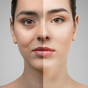
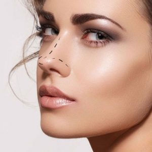
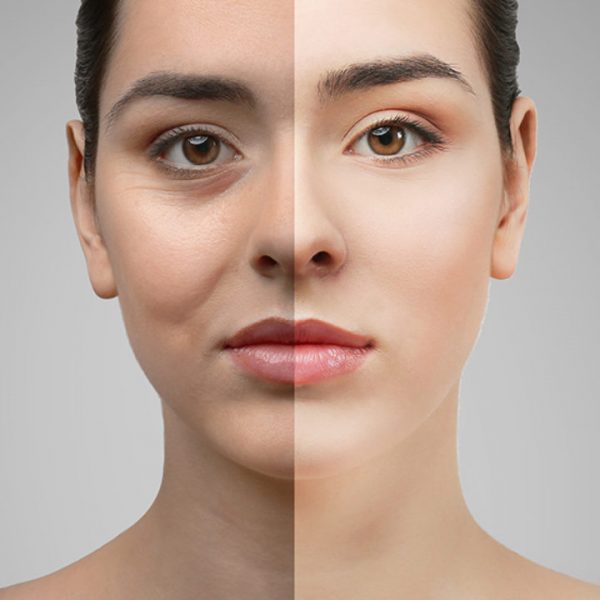
| Content | Blepharoplasty is the general name of the aesthetic corrections we make on the eyelid. Eyelid aesthetics is one of the most common surgical fields of oculoplastic surgery.
Excess skin on the upper eyelid, weakness in the muscle and connective tissue under the skin, collapses in the adipose tissue, sagging of the lacrimal gland; Sagging of the lower eyelid, excess skin, collapsed areas between the lid and cheek, and weakness in the lower lid can be removed by blepharoplasty operations.
The important thing here is to determine which of the above-mentioned procedures will be required by a detailed examination and to plan the surgery that is suitable for the patient meticulously. |
| WOMEN OVER 40 LARGE SCREENING PACKAGE |
| LABORATORY ANALYSIS |
| Glucose |
To determine whether or not your blood glucose level is within normal ranges; to screen for, diagnose, and monitor diabetes, and to monitor for the presence of hypoglycaemia (low blood glucose) and hyperglycaemia (high blood glucose) |
| HbA1c |
To monitor average blood glucose levels over a 3 month period. Used to help diagnose and monitor people with diabetes |
| Urea (Bun) |
To measure how much of waste product you have in your blood. It is used to determine how well your kidneys are working |
| Creatinine |
To assess kidney functions |
| Uric Acid |
To diagnose kidney disorder,diagnose and monitor people with gout, monitor kidney function |
| Complete Urinalysis Test |
To look for metabolic and/or kidney disorders and for urinary tract infections |
| Total Cholesterol |
To screen for risk of developing cardiovascular disease (heart disease, stroke and related diseases); to monitor treatment |
| LDL Cholesterol |
| HDL Cholesterol |
| Triglycerides |
| AST (SGOT) |
To diagnose liver, bile duct and heart diseases |
| ALT (SGPT) |
| GGT |
To screen for liver disease or alcohol abuse; and to help your doctor tell whether a raised concentration of alkaline phosphatase (ALP) in the bloodstream is due to liver or bone disease |
| ALP |
To screen for or monitor treatment for liver or bone disorder |
| Sodium |
To investigate causes of dehydration, oedema, problems with blood pressure, or non-specific symptoms |
| Potassium |
To help diagnose and determine the cause of an electrolyte imbalance; to monitor treatment for illnesses that can cause abnormal potassium levels in the body |
| Chloride |
To determine if there is a problem with your body’s acid-alkali (pH) balance and to monitor treatment |
| Calcium |
To scan, diagnose, and monitor a range of conditions relating to the bones, heart, nerves, kidneys, and teeth |
| Phosphate |
To help in the diagnosis of conditions known to cause abnormally high or low levels |
| Amylase |
To diagnose pancreatitis or other pancreatic diseases |
| Lipase |
To diagnose and monitor pancreatitis or other pancreatic disease |
| Magnesium |
To measure the concentration of magnesium in your blood and to help determine the cause of abnormal calcium and/or potassium levels |
| C-Reactive Protein (CRP) |
To identify the presence of inflammation, to determine its severity, and to monitor response to treatment |
| 25 Hydroxy Vitamin D |
To investigate a problem related to bone metabolism or parathyroid function, possible vitamin D deficiency, malabsorption, before commencing specific bone treatment and to monitor some patients taking vitamin D |
| Blood Count Haemogram |
Haemogram serves as broad screening panel that checks for the presence of any diseases and infections in the body |
| Erythrocyte Sedimentation Rate
(ESR) |
To detect and monitor the activity of inflammation as an aid in the diagnosis of the underlying cause |
| Ferritine |
To help assess the levels of iron stored in your body |
| Vitamin B12 |
To help diagnose the cause of anaemia or neuropathy (nerve damage), to evaluate nutritional status in some patients, to monitor effectiveness of treatment of B12 or folate deficiency |
| Free T3 |
To help diagnose hyperthyroidism and monitor it's treatment |
| Free T4 |
To diagnose hypothyroidism or hyperthyroidism in adults and to monitor response to treatment |
| TSH |
To screen for and diagnose thyroid disorders; to monitor treatment of hypothyroidism and hyperthyroidism |
| FSH |
To evaluate the function of your pituitary gland, which regulates the hormones that control your reproductive system |
| LH |
| Estrogens (E2) |
To measure or monitor your estrogen levels; to detect an abnormal level or hormone imbalance ; to monitor treatment for infertility or symptoms of menopause; sometimes to test for fetal-placental status during early stages of pregnancy |
| Prolactin |
To diagnose a prolactinoma (a type of tumor of the pituitary gland, help tofind the cause of a woman's menstrual irregularities and/or infertility. Or to help to find the cause of a man's low sex drive and/or erectile dysfunction |
| Beta hCG |
To confirm and monitor pregnancy or to diagnose trophoblastic disease or germ cell tumours |
| HBsAg |
To detect, diagnose and follow the course of an infection with hepatitis B virus (HBV) or to determine if the vaccine against hepatitis B has produced the desired level of immunity |
| Anti HBs |
| Anti HCV |
To screen for and diagnose hepatitis C virus infection and to monitor treatment of the infection |
| Anti HIV |
To determine if you are infected with human immunodeficiency virus (HIV) |
| CEA |
In the presence of certain cancers, CEA may be used to monitor the effect of treatment and recurrence of disease |
| CA 125 |
To monitor treatment for ovarian cancer or to investigate for a possible ovarian cancer |
| CA 19-9 |
To help tell the difference between cancer of the pancreas and bile ducts and other conditions; to monitor response to pancreatic cancer treatment and to watch for recurrence |
| CA 15-3 |
To monitor the response to treatment of breast cancer and to watch for recurrence of the disease |
| AFP |
To screen for and monitor therapy for certain cancers of the liver and testes |
| Fecal Direct Parasite Search (Ova
& Parasite Exam) |
To determine whether you have a parasite infecting your digestive tract |
| Fecal Occult Blood Test |
To screen for bleeding from the gut/intestine, which may be an indicator of bowel cancer |
| OTHER ANALYSIS |
| Abdominal Ultrasound |
To identify diseases at organs in the abdomen, including the liver, gallbladder, spleen, pancreas, and kidneys |
| Thyroid Ultrasound |
To characterize a thyroid nodule(s), i.e. to measure the dimensions accurately and to identify internal structure and vascularization |
| Carotid Ultrasound |
To detect narrowing, or stenosis, of the carotid artery, a condition that substantially increases the risk of stroke |
| Breast Ultrasound (Bilateral) |
To screen suspected breast cancer or for early diagnosis and control. It is the imaging of breast with ultrasound device |
| Mammography (Bilateral) |
| Pap Smear |
Method for early diagnosis of Cervical cancer and infectious diseases |
| Echocardiogram |
To evaluate how your heart moves, heart valves are working and heart’s pumping strength |
| Electrocardiogram |
To measure the electrical activity of the heartbeat and hearth rhythm |
| Exercise Stress Test |
To determine how well your hearth handles work. The test can show if the blood supply is reduced in the arteries that supply the heart |
| Pulmonary Function Test |
To tests that measure how well your lungs work. |
| Chest X-Ray |
The most commonly preferred diagnostic examination to produce images of heart, lungs, airways, blood vessels and the bones of the spine and chest |
| EXAMINATIONS |
| Internal Medicine Examination |
General physical examination, evaluation of the results and recommendations |
| Cardiology Examination |
| Gynaecology Examination |
| Ophtalmology Examination |
| Pulmonology Examination |
| General Surgery Examination |
|
|
|
MEN OVER 40 LARGE SCREENING PACKAGE
|
| LABORATORY ANALYSIS
|
| Glucose |
To determine whether or not your blood glucose level is within normal ranges; to screen for, diagnose, and monitor diabetes, and to monitor for the presence of hypoglycaemia (low blood glucose) and hyperglycaemia (high blood glucose) |
| HbA1c |
To monitor average blood glucose levels over a 3 month period. Used to help diagnose and monitor people with diabetes |
| Urea (Bun) |
To measure how much of waste product you have in your blood. It is used to determine how well your kidneys are working |
| Creatinine |
To assess kidney functions |
| Uric Acid |
To diagnose kidney disorder,diagnose and monitor people with gout, monitor kidney function |
| Complete Urinalysis Test |
To look for metabolic and/or kidney disorders and for urinary tract infections |
| Total Cholesterol |
To screen for risk of developing cardiovascular disease (heart disease, stroke and related diseases); to monitor treatment |
| LDL Cholesterol |
| HDL Cholesterol |
| Triglycerides |
| AST (SGOT) |
To diagnose liver, bile duct and heart diseases |
| ALT (SGPT) |
| GGT |
To screen for liver disease or alcohol abuse; and to help your doctor tell whether a raised concentration of alkaline phosphatase (ALP) in the bloodstream is due to liver or bone disease |
| ALP |
To screen for or monitor treatment for liver or bone disorder |
| Sodium |
To investigate causes of dehydration, oedema, problems with blood pressure, or non-specific symptoms |
| Potassium |
To help diagnose and determine the cause of an electrolyte imbalance; to monitor treatment for illnesses that can cause abnormal potassium levels in the body |
| Chloride |
To determine if there is a problem with your body’s acid-alkali (pH) balance and to monitor treatment |
| Calcium |
To scan, diagnose, and monitor a range of conditions relating to the bones, heart, nerves, kidneys, and teeth |
| Phosphate |
To help in the diagnosis of conditions known to cause abnormally high or low levels |
| Amylase |
To diagnose pancreatitis or other pancreatic diseases |
| Lipase |
To diagnose and monitor pancreatitis or other pancreatic disease |
| Magnesium |
To measure the concentration of magnesium in your blood and to help determine the cause of abnormal calcium and/or potassium levels |
| C-Reactive Protein (CRP) |
To identify the presence of inflammation, to determine its severity, and to monitor response to treatment |
| 25 Hydroxy Vitamin D |
To investigate a problem related to bone metabolism or parathyroid function, possible vitamin D deficiency, malabsorption, before commencing specific bone treatment and to monitor some patients taking vitamin D |
| Blood Count Haemogram |
Haemogram serves as broad screening panel that checks for the presence of any diseases and infections in the body |
| Erythrocyte Sedimentation Rate
(ESR) |
To detect and monitor the activity of inflammation as an aid in the diagnosis of the underlying cause |
| Ferritine |
To help assess the levels of iron stored in your body |
| Vitamin B12 |
To help diagnose the cause of anaemia or neuropathy (nerve damage), to evaluate nutritional status in some patients, to monitor effectiveness of treatment of B12 or folate deficiency |
| Free T3 |
To help diagnose hyperthyroidism and monitor it's treatment |
| Free T4 |
To diagnose hypothyroidism or hyperthyroidism in adults and to monitor response to treatment |
| TSH |
To screen for and diagnose thyroid disorders; to monitor treatment of hypothyroidism and hyperthyroidism |
| Beta hCG |
To confirm and monitor pregnancy or to diagnose trophoblastic disease or germ cell tumours |
| HBsAg |
To detect, diagnose and follow the course of an infection with hepatitis B virus (HBV) or to determine if the vaccine against hepatitis B has produced the desired level of immunity |
| Anti HBs |
| Anti HCV |
To screen for and diagnose hepatitis C virus infection and to monitor treatment of the infection |
| Anti HIV |
To determine if you are infected with human immunodeficiency virus (HIV) |
| CEA |
In the presence of certain cancers, CEA may be used to monitor the effect of treatment and recurrence of disease |
| CA 19-9 |
To help tell the difference between cancer of the pancreas and bile ducts and other conditions; to monitor response to pancreatic cancer treatment and to watch for recurrence |
| AFP |
To screen for and monitor therapy for certain cancers of the liver and testes |
| Total PSA |
To help detect and to monitor prostate cancer |
| Free PSA |
| Fecal Direct Parasite Search
(Ova & Parasite Exam) |
To determine whether you have a parasite infecting your digestive tract |
| Fecal Occult Blood Test |
To screen for bleeding from the gut/intestine, which may be an indicator of bowel cancer |
| Abdominal Ultrasound |
To identify diseases at organs in the abdomen, including the liver, gallbladder, spleen, pancreas, and kidneys |
| Thyroid Ultrasound |
To characterize a thyroid nodule(s), i.e. to measure the dimensions accurately and to identify internal structure and vascularization |
| Carotid Ultrasound |
To detect narrowing, or stenosis, of the carotid artery, a condition that substantially increases the risk of stroke |
| Echocardiogram |
To evaluate how your heart moves, heart valves are working and heart’s pumping strength |
| Electrocardiogram |
To measure the electrical activity of the heartbeat and hearth rhythm |
| Exercise Stress Test |
To determine how well your hearth handles work. The test can show if the blood supply is reduced in the arteries that supply the heart |
| Pulmonary Function Test |
To tests that measure how well your lungs work. |
| Chest X-Ray |
The most commonly preferred diagnostic examination to produce images of heart, lungs, airways, blood vessels and the bones of the spine and chest |
| EXAMINATIONS |
| Internal Medicine Examination |
General physical examination, evaluation of the results and recommendations |
| Cardiology Examination |
| Ophtalmology Examination |
| Pulmonology Examination |
| Urology Examination |
| General Surgery Examination |
| Dermatology Examination |
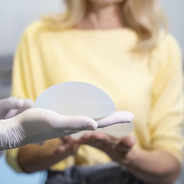
| Big enough and firm breasts for a woman both increase self-confidence and create great freedom in choosing clothes. However, most women do not have ideal measurements. Especially women with small breasts or those with no breasts feel the most inadequate and unhappy. |
| GENERAL CARDIOLOGY SCREENING PACKAGE |
| LABORATORY ANALYSIS |
| Glucose |
To determine whether or not your blood glucose level is within normal ranges; to screen for, diagnose, and monitor diabetes, and to monitor for the presence of hypoglycaemia (low blood glucose) and hyperglycaemia (high blood glucose) |
| HbA1c |
To monitor average blood glucose levels over a 3 month period. Used to help diagnose and monitor people with diabetes |
| Urea (Bun) |
To measure how much of waste product you have in your blood. It is used to determine how well your kidneys are working |
| Creatinine |
To assess kidney functions |
| Uric Acid |
To diagnose kidney disorder,diagnose and monitor people with gout, monitor kidney function |
| Complete Urinalysis Test |
To look for metabolic and/or kidney disorders and for urinary tract infections |
| Total Cholesterol |
To screen for risk of developing cardiovascular disease (heart disease, stroke and related diseases); to monitor treatment |
| LDL Cholesterol |
| HDL Cholesterol |
| Triglycerides |
| AST (SGOT) |
To diagnose liver, bile duct and heart diseases |
| ALT (SGPT) |
| Sodium |
To investigate causes of dehydration, oedema, problems with blood pressure, or non-specific symptoms |
| Potassium |
To help diagnose and determine the cause of an electrolyte imbalance; to monitor treatment for illnesses that can cause abnormal potassium levels in the body |
| Calcium |
To scan, diagnose, and monitor a range of conditions relating to the bones, heart, nerves, kidneys, and teeth |
| C-Reactive Protein (CRP) |
To identify the presence of inflammation, to determine its severity, and to monitor response to treatment |
| 25 Hydroxy Vitamin D |
To investigate a problem related to bone metabolism or parathyroid function, possible vitamin D deficiency, malabsorption, before commencing specific bone treatment and to monitor some patients taking vitamin D |
| Homocysteine |
To find out if you are at high risk of a heart attack or stroke; also used to determine if you are folate-deficient or vitamin B12-deficient |
| Lipoprotein A |
To evaluate targeted screening for cardiovascular disease (coronary artery disease (CAD) and cerebrovascular disease) risk assessment |
| Blood Count Haemogram |
Haemogram serves as broad screening panel that checks for the presence of any diseases and infections in the body. |
| Troponin |
To see if you have had a heart attack or damage to your heart muscle |
| Vitamin B12 |
To help diagnose the cause of anaemia or neuropathy (nerve damage), to evaluate nutritional status in some
patients, to monitor effectiveness of treatment of B12 or folate deficiency |
| Free T3 |
To help diagnose hyperthyroidism and monitor it's treatment |
| Free T4 |
To diagnose hypothyroidism or hyperthyroidism in adults and to monitor response to treatment |
| TSH |
To screen for and diagnose thyroid disorders; to monitor treatment of hypothyroidism and hyperthyroidism |
| D- dimer |
To help diagnose or exclude thrombotic (blood clot producing) or bleeding diseases and conditions |
| OTHER ANALYSIS |
| Carotid Ultrasound |
To detect narrowing, or stenosis, of the carotid artery, a condition that substantially increases the risk of stroke |
| Echocardiogram |
To evaluate how your heart moves, heart valves are working and heart’s pumping strength |
| Electrocardiogram |
To measure the electrical activity of the heartbeat and hearth rhythm |
| Exercise Stress Test |
To determine how well your hearth handles work. The test can show if the blood supply is reduced in the arteries that supply the heart |
| EXAMINATIONS |
| Cardiology Examination |
General physical examination, evaluation of the results and recommendations |
| General Surgery Examination |
| Dermatology Examination |
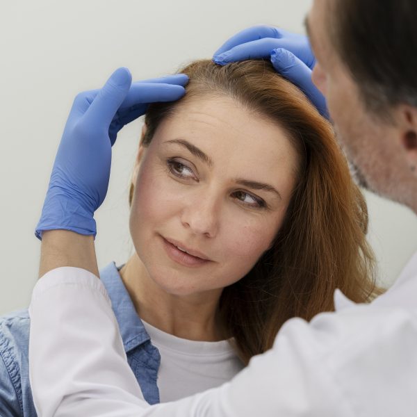
| Hair loss, which is one of the biggest problems of today, has become a problem that women encounter more frequently than men. Yaşam Hospital hair transplantation department is a center that follows world standards in hair transplantation and works with professional experienced staff in this field. It follows the scientific developments related to hair transplantation and serves its patients with the latest technology. Our center is a full-fledged hospital, and our procedures are carried out in a sterile environment with care for you and your health. |










Reviews
There are no reviews yet.