Online diet eliminates space and accessibility problems. Wherever you are in the world, your dietitian is always with you wherever there is internet.
With an online diet, your time is up to you. If you cannot find time to go to a dietitian for reasons such as busy work life, child care, traffic problems in big cities, you can meet with your dietitian at home, on your business trips, or anywhere you go to spend time with your family, thanks to the online diet.
It is much easier to follow a dietitian in the online diet system. It is extremely important that your dietitian follows you throughout your diet program. In the online diet system, your dietitian can follow you much more closely.
The sustainability rate of the online diet is much higher. Sometimes it is not possible to go to a dietitian regularly and clients leave their diet programs unfinished. However, you do not have to make time to go to a dietitian in an online diet.
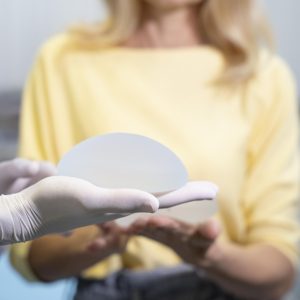
Breast Health Package for Women Over 40
Warning: call_user_func_array() expects parameter 1 to be a valid callback, function 'digic_get_brands' not found or invalid function name in /home/antalyac/public_html/wp-includes/class-wp-hook.php on line 324
€99,00
Warning: call_user_func_array() expects parameter 1 to be a valid callback, function 'digic_show_store_name' not found or invalid function name in /home/antalyac/public_html/wp-includes/class-wp-hook.php on line 324
| Select Hospital | Antalya Yaşam Hospital, Kemer Yaşam Hospital, ASV Yaşam Hospital, Opera Yaşam Hospital, Alanya Yaşam Hospital, Manavgat Yaşam Hospital |
|---|
You must be logged in to post a review.
Vendor Information
- Address:
- No ratings found yet!
-
Warning: call_user_func_array() expects parameter 1 to be a valid callback, function 'digic_woocommerce_template_loop_category' not found or invalid function name in /home/antalyac/public_html/wp-includes/class-wp-hook.php on line 324
Home Care
€280,00 -
Warning: call_user_func_array() expects parameter 1 to be a valid callback, function 'digic_woocommerce_template_loop_category' not found or invalid function name in /home/antalyac/public_html/wp-includes/class-wp-hook.php on line 324
Men Under 40 Large Screening Package
€680,00 -
Warning: call_user_func_array() expects parameter 1 to be a valid callback, function 'digic_woocommerce_template_loop_category' not found or invalid function name in /home/antalyac/public_html/wp-includes/class-wp-hook.php on line 324
Inpatient Men Vip Check-Up
€2.250,00 -
Warning: call_user_func_array() expects parameter 1 to be a valid callback, function 'digic_woocommerce_template_loop_category' not found or invalid function name in /home/antalyac/public_html/wp-includes/class-wp-hook.php on line 324
Hair Transplant
€1.750,00 -
Warning: call_user_func_array() expects parameter 1 to be a valid callback, function 'digic_woocommerce_template_loop_category' not found or invalid function name in /home/antalyac/public_html/wp-includes/class-wp-hook.php on line 324
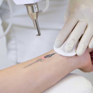
Tattoo Removal
€170,00 -
Warning: call_user_func_array() expects parameter 1 to be a valid callback, function 'digic_woocommerce_template_loop_category' not found or invalid function name in /home/antalyac/public_html/wp-includes/class-wp-hook.php on line 324
Neural Therapy
€30,00
Quick Comparison
| Settings | Breast Health Package for Women Over 40 remove | Mesotherapy In Hair Loss remove | Abdominoplasty remove | Online Diet remove | Hair Transplant remove | Vaginoplasty remove | ||||||||
|---|---|---|---|---|---|---|---|---|---|---|---|---|---|---|
| Name | Breast Health Package for Women Over 40 remove | Mesotherapy In Hair Loss remove | Abdominoplasty remove | Online Diet remove | Hair Transplant remove | Vaginoplasty remove | ||||||||
| Image |  | 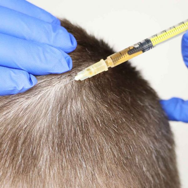 |  |  | 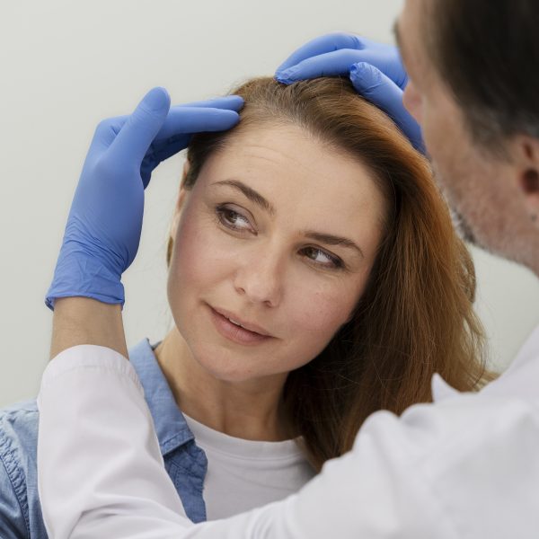 |  | ||||||||
| SKU | D2300-3-2-3-2-1-1-1-1-2-2-1-3 | |||||||||||||
| Rating | ||||||||||||||
| Price | €99,00 | €175,00 | €3.450,00 | €60,00 | €1.750,00 | €1.700,00 | ||||||||
| Stock | ||||||||||||||
| Availability | ||||||||||||||
| Add to cart | ||||||||||||||
| Description | Abdominal stretching surgeries vary according to the technique applied and the severity of the patient's complaint. Which one should be applied to you is decided after the pre-operative interview and examination. | Our hair purchases are made with the FUE (Follicular Unit Extraction) method. FUE is the process of removing hair islands (follicular unit; FU) one by one without making a linear incision with the help of a micromotor. Within 1-2 weeks after the procedure, visible improvement is completed in the area where the procedure is taken. Daily life can be easily continued. Our plantings are state-of-the-art as well as the classical canal method. It is done with the DHI method (pencil method), which gives the healthiest and most natural results. | ||||||||||||
| Content |
Breast Health Package for Women Over 40Our Breast Health Center, which is a part of Yaşam Hospital Oncology Center, offers all the possibilities of technology to provide the best care to every woman. What is Mammography?Mammography is the low-dose X-ray imaging of the breast tissue to look for early signs of breast cancer before symptoms develop. It can also be used for due diligence when a new symptom (lump or focal pain) develops in the breast tissue. When viewed on a mammogram, breast tissue appears white and opaque (nebula), while fatty tissue appears darker and translucent.When Should a Mammogram Be Done?Annual screening mammograms are recommended for all women from the age of 40. Women who do not have any breast-related signs or symptoms are also screened. If an abnormality is present or patients have a new symptom (a lump or focal pain), additional evaluation may be required. Further examination will reveal what these suspected abnormalities are.What Percent of Women Who Have Had Mammography Have Risky Situations?Potential abnormalities are found in 6 to 8 percent of women who get mammograms. This group undergoes different additional evaluations, which may include breast physical examination, diagnostic mammography, breast ultrasound or needle biopsy.After these additional evaluations are completed, it becomes clear what the abnormalities found on the mammogram are.What Does an Abnormality Look Like on a Mammogram?The possible abnormality on a mammogram may be called a nodule, mass, lump, density or deterioration: A mass (lump) with a smooth, well-defined border is usually benign. Ultrasound is necessary to see and identify the inside of a mass. If the mass contains fluid, it is called a cyst. A mass (lump) with irregular borders or a starburst appearance may be cancerous and a biopsy is usually recommended. Microcalcifications (small calcium deposits) are another type of abnormality. They can be classified as benign, suspicious, or uncertain. Most microcalcifications are benign. Depending on how the microcalcifications appear in additional studies (magnification views), a biopsy may be recommended.What is the Accuracy Rate of Mammography?Diagnoses made by mammography are between 85 percent and 90 percent accurate. Mammograms can detect breast abnormalities before they are large enough to be felt. However, a palpable mass may not be seen on a mammogram. Any abnormality you feel while examining your breasts should be evaluated by your doctor.What Should Be Considered Before Mammography?You can follow your normal routine before the mammogram. You can take your medications and maintain your eating and drinking patterns. If you are breastfeeding, pregnant or think you may be pregnant, you should tell your doctor as your mammogram may need to be postponed.What Should I Pay Attention to When Coming to My Mammography Appointment?The technician will ask you to remove one breast from your bib at a time and place it on the chest support plate. The image of the breast is taken by clamping it between two plates. In the meantime, pressure is applied to the breast, preventing the breast from moving. This pressure spreads the breast tissue, allowing the radiologist to see the tissue better. Also, the least amount of radiation is used when the breast is compressed as finely as possible. You may feel some discomfort during 3-5 seconds of pressure. If you cannot tolerate the pressure, please let the technician know. Pressure can be more bothersome at some times in a woman’s menstrual cycle. To minimize discomfort, we recommend scheduling your appointment seven to 10 days after the start of your period.How is a Mammogram Taken?The technician will ask you to remove one breast from your bib at a time and place it on the chest support plate. The image of the breast is taken by clamping it between two plates. In the meantime, pressure is applied to the breast, preventing the breast from moving. This pressure spreads the breast tissue, allowing the radiologist to see the tissue better. Also, the least amount of radiation is used when the breast is compressed as finely as possible. You may feel some discomfort during 3-5 seconds of pressure. If you cannot tolerate the pressure, please let the technician know. Pressure can be more bothersome at some times in a woman’s menstrual cycle. To minimize discomfort, we recommend scheduling your appointment seven to 10 days after the start of your period.How Does the Process Proceed After Mammography?There may be temporary skin discoloration and/or mild pain in the chest due to compression. Most women will be able to resume their normal activities soon after their mammogram. Your results will be available within a few days after the test. After getting the results, your doctor will explain everything to you.How Often Should You Have a Mammogram?Regular mammograms every year, starting at the age of 40, will enable you to recognize potential risks early.What Does the Breast Health Package Consist of?Breast health package consists of General Surgery Examination and Mammography | With hair mesotherapy, vitamines and minerals which immediately affect hair growth, are directly applied to the hair root. Hair mesotherapy is used for all kinds of hair loss(man-type hair loss, hormonal hair loss, anemia etc.). Asthe therapy is made with very thin needles, it doesn’t hurt very much. Results start to be visible after the 3rd session. After a total of 8-10 sessions, a recall session should be applied after 3-4 months. | During pregnancy, due to sudden growth in the abdomen or as a result of weight gain or loss, sagging in the abdomen, cracks, etc. There are some undesirable changes. Abdominal stretching helps to reduce these complaints. In fact, the patients with the best results are those who have come because of their sagging after weight loss. Although it is not possible for all the cracks on the abdomen to disappear in tummy tuck surgeries, it is possible to reduce them even more. There is no doubt that it is appropriate to perform this type of surgery by Aesthetic and Plastic surgery specialists, in appropriate operating room conditions and when all conditions that protect the patient are met. | Hair loss, which is one of the biggest problems of today, has become a problem that women encounter more frequently than men. Yaşam Hospital hair transplantation department is a center that follows world standards in hair transplantation and works with professional experienced staff in this field. It follows the scientific developments related to hair transplantation and serves its patients with the latest technology. Our center is a full-fledged hospital, and our procedures are carried out in a sterile environment with care for you and your health. |
VaginoplastyVaginoplasty is a procedure to repair the vagina. It treats a variety of medical issues, including vaginal enlargements from childbirth and complications of pelvic floor disease.What is done during vaginoplasty?The details of the procedure vary depending on your goals and medical needs. The vagina is reconstructed using various surgical techniques.Who needs vaginoplasty?
What is the difference between vaginoplasty and other vaginal operations?
What is done before vaginoplasty?
What is the content of the procedure in women with postpartum deformation?
What is the content of vaginoplasty to repair congenital (birth) defects?
Risks / Benefits
What is the recovery process like after vaginoplasty?
How are the controls planned after vaginoplasty?
| |||||||||
| Weight | N/A | N/A | N/A | N/A | N/A | N/A | ||||||||
| Dimensions | N/A | N/A | N/A | N/A | N/A | N/A | ||||||||
| Additional information |
|
|
|
|








Reviews
There are no reviews yet.