| Content |
Breast Health Package for Women Under 40
Our Breast Health Center, which is a part of Yaşam Hospital Oncology Center, offers all the possibilities of technology to provide the best care to every woman.
Early Detection of Breast Cancer
Breast cancer is the most common type of cancer that women can encounter in their lifetime (Early detection can save your life).
Self Examination
From the age of 20, all women should perform a breast self-exam once a month (about one week after the start of each menstrual period). If you feel an unusual lump or anything else, it’s important to contact your doctor right away.
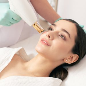
What is Breast Ultrasound?
Ultrasound is an imaging method that uses high-frequency sound waves to take pictures of internal organs and tissues.
When Should Breast Ultrasound Be Done?
From the age of 25, annual routine ultrasound follow-ups should not be interrupted. Ultrasound can be applied safely as it does not contain radiation and has no side effects on the body.
What Are the Risks of Breast Ultrasound?
Breast ultrasound uses sound waves, not radiation, to produce images. Ultrasound technology has no known risks.
What Happens After Breast Ultrasound?
After your breast ultrasound, the radiologist interprets the images and reports the results to your doctor. This information will also be shared with you if any additional testing is needed or follow-up is recommended.
What are the Benefits of Breast Ultrasound?
Ultrasound scanning is non-invasive (no needles or injections).
Ultrasound imaging is extremely safe and uses no radiation.
An ultrasound scan gives a clear picture of soft tissues that don’t show up well on x-ray images.
Ultrasound provides real-time imaging. This makes it easy to guide minimally invasive procedures such as needle biopsies and fluid aspiration.
Ultrasound imaging helps detect lesions in women with dense breasts.
Ultrasound can help detect and classify a breast lesion that cannot be adequately interpreted by mammography alone.
Using ultrasound, doctors can determine that many areas are caused by normal tissue (such as fat lobules) or benign cysts. For most women age 40 and older, a mammogram will be used along with the ultrasound. For women under the age of 40, ultrasound alone is often sufficient to determine whether an area of concern needs a biopsy.
What Does the Breast Health Package Consist of?
Breast health package consists of General Surgery Examination and Breast USG.
Note from your doctor:
Breast ultrasound is a safe, painless imaging method for examining targeted areas of breast tissue. With breast ultrasound, we provide detailed images of breast tissue and can diagnose cysts or lumps if present. Diagnosing possible risks at an early stage provides a very important gain in the treatment process. |
Labiaplasty Package
Labiaplasty is a surgical procedure to reduce or enlarge the skin folds that usually surround your labia and vagina. Excess skin can be bent and pinched, causing discomfort during exercise, physical activities, and sexual intercourse.
Depending on why the procedure is performed during labiaplasty, your doctor may:
It removes some tissue from the labia to reduce its size.
Injects filler or oil to enlarge the labia.
It forms a labia from other tissues.
What are the reasons for requesting labiaplasty?
Reducing labia minora size
Excess lip tissue can be bent, twisted, compressed. This, can cause physical discomfort and irritation during exercise, physical activities (such as cycling or jogging), and sexual intercourse.
Reducing the size of the labia minora may be desirable to improve hygiene and health, as excess tissue can make cleaning difficult and may harbor bacteria that can lead to the development of urinary tract infections.
Cosmetic and emotional reasons
To have a younger appearance after birth or aging,
Reducing asymmetry of the labia minora or labia majora when one side is longer or has a different shape than the other,
To increase self-confidence and to eliminate visual lines and protrusions when wearing body-fitting trousers or tights,
During close contact, labiaplasty can be performed to improve comfort, confidence and sexual health regarding the appearance of your genitals.
What causes an overgrowth of a labia?
Age, menopause, or other hormonal changes can thin the labia majora tissue, causing the labia minora to protrude from the labia majora.
Pregnancy and childbirth.
Changes in your weight.
Genetic.
What are the pre-operative procedures?
First, you and your doctor will discuss why you want to have labiaplasty and make sure that the surgery will meet your expectations. During the physical examination, your doctor will explain where the incisions will be made and what to expect regarding changes in the size and shape of your labia.
After the surgery decision, your doctor will inform you in detail about the preparation processes for the surgery. In this process, your doctor will inform you about how to resize or reshape your labia (labia majora and/or labia minora) and will enlighten you about the surgical procedure to be applied.
What is the procedure after labiaplasty surgery?
After the surgery, your doctor will give you the necessary information on how to care. Carefully applying the information conveyed to you about what you should and shouldn’t do during the recovery process will speed up your recovery process. We also recommend that you make your control appointments before leaving the hospital.
What are the risks and complications of labiaplasty surgery?
- - Complications of laboplasty surgery are rare. Even a little;
- - Bleeding,
- - Bruising, swelling (hematoma),
- - Infection,
- - Scar,
- - Ongoing pain, pain with sexual intercourse, or loss of sensitivity may occur.
- - In these cases, you can consult your doctor.
Is the recovery process painful after labiaplasty?
- It is normal to feel some swelling, discomfort and pain after surgery. Most people report only mild pain or tenderness for a few days. In return, your doctor will advise you on medication for your pain.Wearing loose trousers and underwear during the recovery period will accelerate your healing process.
Will it bleed a lot?
You may have a small amount of bleeding for a week. This is normal, you can use pads to absorb the blood.
When will the satisfactory results of labiaplasty be obtained?
Initial swelling, pain, and temporary discoloration subside within a few weeks after surgery. However, you may have to wait up to four to six months to see the final results of your labiaplasty. Post-operative scarring is usually negligible.
How do I wash my labia area?
Use only warm water to wash your wound (do not use soap) and wipe gently, do not rub the surgical site.
It is important to follow your doctor’s instructions to keep the surgical site clean and free of bacteria and to speed the healing of the surgical site.
When can I return to work, physical activity and sex life?
You should be able to return to work and other light activities after a few days. If your job involves intense physical activity or lifting weights, you can get more detailed information by informing your doctor about this. until your doctor approves. |
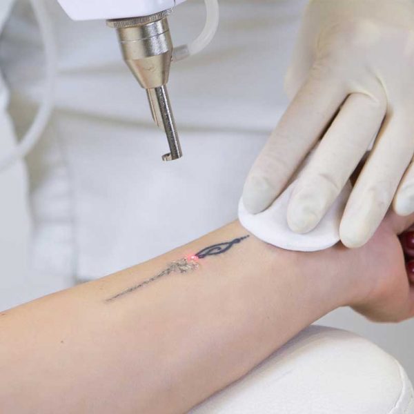
What is laser tattoo removal?
Tattooing is a permanent procedure on the skin. Tattoo removal can be done with modern methods when, for any reason, the person does not want to see his tattoo on his body anymore.
Tattoo removal, permanent make-up and stain treatment can be performed with the Q-switch Nd:YAG laser. This process is a technology that destroys the color pigments used in tattoos by focusing. During this very delicate process, the laser sees and destroys the pigment and damages the tissue as little as possible. In laser tattoo removal, which is a long and laborious process, one hundred percent result may not always be achieved. For this, the decision to have a tattoo should be considered very well, and possible future regrets should be considered.
How is laser tattoo removal done?
Tattoo and skin analysis should be done before the procedure. Lasers designed for tattoo removal and spot treatment send the beam to the epidermis layer of the skin, absorb the compounds that give color to the dermis and subcutaneous tattoo, and destroy the chemical substance by breaking it down. The wavelength of the laser; is adjusted according to the size, density and color of the tattoo.
In laser shots, dye pigments are made without damaging the tissues.
While black dyes under the skin are better detected by the laser, more effective results are obtained, while the same is not true for light-colored dyes.
Different wave sizes are used in tattoos with multi-colored paints.
How many sessions does laser tattoo removal take?
There are some environmental and structural factors in tattoo removal. It is important in which part of the body the tattoo is located. More sessions are required to remove tattoos, especially on the tips of the feet and fingers. For tattoos on larger areas of the body, a more effective process is performed and removed. The duration of the erasing process varies due to the color of the tattoo, the amount and type of paint used in the tattoo. Tattoo removal sessions take place at intervals of approximately 1.5 – 2 months. The number of sessions varies according to the person and the nature of the tattoo.
Will there be any scars after laser tattoo removal?
After each tattoo removal session, the color of the tattoo will lighten by approximately 20%. In tattoos where multi-colored and phosphorescent colors are used, the erasing process may be longer.
Is laser tattoo removal painful?
After numbing the skin part with cream or local anesthesia, the procedure is performed by protecting the upper skin layer in that area with the help of a Q-Switch ND-YAG laser and cooler. After the procedure, there may be bleeding in the area and crusting on the skin that will last for about a few days after the procedure. After the application, antibiotic cream should be used to both repair the area and prevent the development of infection.
When should laser tattoo removal be done?
While tattoo removal is more comfortable in winter, it is not preferred in summer due to the risk of staining with the effect of the sun. The time of the procedure varies according to the size of the tattoo. Since the sun is out in the summer, problems such as permanent staining may occur after the procedure. The best time for this is the winter months. |
| MEN UNDER 40 LARGE SCREENING PACKAGE |
| Glucose |
To determine whether or not your blood glucose level is within normal ranges; to screen for, diagnose, and monitor diabetes, and to monitor for the presence of hypoglycaemia (low blood glucose) and hyperglycaemia (high blood glucose) |
| HbA1c |
To monitor average blood glucose levels over a 3 month period. Used to help diagnose and monitor people with diabetes |
| Urea (Bun) |
To measure how much of waste product you have in your blood. It is used to determine how well your kidneys are working |
| Creatinine |
To assess kidney functions |
| Uric Acid |
To diagnose kidney disorder,diagnose and monitor people with gout, monitor kidney function |
| Complete Urinalysis Test |
To look for metabolic and/or kidney disorders and for urinary tract infections |
| Total Cholesterol |
To screen for risk of developing cardiovascular disease (heart disease, stroke and related diseases); to monitor treatment |
| LDL Cholesterol |
| HDL Cholesterol |
| Triglycerides |
| AST (SGOT) |
To diagnose liver, bile duct and heart diseases |
| ALT (SGPT) |
| GGT |
To screen for liver disease or alcohol abuse; and to help your doctor tell whether a raised concentration of alkaline phosphatase (ALP) in the bloodstream is due to liver or bone disease |
| ALP |
To screen for or monitor treatment for liver or bone disorder |
| Sodium |
To investigate causes of dehydration, oedema, problems with blood pressure, or non-specific symptoms |
| Potassium |
To help diagnose and determine the cause of an electrolyte imbalance; to monitor treatment for illnesses that can cause abnormal potassium levels in the body |
| Chloride |
To determine if there is a problem with your body’s acid-alkali (pH) balance and to monitor treatment |
| Calcium |
To scan, diagnose, and monitor a range of conditions relating to the bones, heart, nerves, kidneys, and teeth. |
| Phosphate |
To help in the diagnosis of conditions known to cause abnormally high or low levels |
| Amylase |
To diagnose pancreatitis or other pancreatic diseases |
| Lipase |
To diagnose and monitor pancreatitis or other pancreatic disease |
| Magnesium |
To measure the concentration of magnesium in your blood and to help determine the cause of abnormal calcium and/or potassium levels |
| C-Reactive Protein (CRP) |
To identify the presence of inflammation, to determine its severity, and to monitor response to treatment |
| 25 Hydroxy Vitamin D |
To investigate a problem related to bone metabolism or parathyroid function, possible vitamin D deficiency, malabsorption, before commencing specific bone treatment and to monitor some patients taking vitamin D |
| Blood Count Haemogram |
Haemogram serves as broad screening panel that checks for the presence of any diseases and infections in the body |
| Erythrocyte Sedimentation Rate
(ESR) |
To detect and monitor the activity of inflammation as an aid in the diagnosis of the underlying cause |
| Ferritine |
To help assess the levels of iron stored in your body |
| Vitamin B12 |
To help diagnose the cause of anaemia or neuropathy (nerve damage), to evaluate nutritional status in some patients, to monitor effectiveness of treatment of B12 or folate deficiency |
| Free T3 |
To help diagnose hyperthyroidism and monitor it's treatment |
| Free T4 |
To diagnose hypothyroidism or hyperthyroidism in adults and to monitor response to treatment |
| TSH |
To screen for and diagnose thyroid disorders; to monitor treatment of hypothyroidism and hyperthyroidism |
| HBsAg |
To detect, diagnose and follow the course of an infection with hepatitis B virus (HBV) or to determine if the vaccine against hepatitis B has produced the desired level of immunity |
| Anti HBs |
| Anti HCV |
To screen for and diagnose hepatitis C virus infection and to monitor treatment of the infection |
| Anti HIV |
To determine if you are infected with human immunodeficiency virus (HIV) |
| Fecal Occult Blood Test |
To screen for bleeding from the gut/intestine, which may be an indicator of bowel cancer |
| OTHER ANALYSIS |
| Abdominal Ultrasound |
To identify diseases at organs in the abdomen, including the liver, gallbladder, spleen, pancreas, and kidneys |
| Thyroid Ultrasound |
To characterize a thyroid nodule(s), i.e. to measure the dimensions accurately and to identify internal structure and vascularization |
| Echocardiogram |
To evaluate how your heart moves, heart valves are working and heart’s pumping strength |
| Electrocardiogram |
To measure the electrical activity of the heartbeat and hearth rhythm |
| Exercise Stress Test |
To determine how well your hearth handles work. The test can show if the blood supply is reduced in the arteries that supply the heart |
| Pulmonary Function Test |
To tests that measure how well your lungs work. |
| Chest X-Ray |
The most commonly preferred diagnostic examination to produce images of heart, lungs, airways, blood vessels and the bones of the spine and chest |
| EXAMINATIONS |
| Internal Medicine Examination |
General physical examination, evaluation of the results and recommendations |
| Cardiology Examination |
| Ophtalmology Examination |
| Pulmonology Examination |
| Urology Examination |
| General Surgery Examination |
| Dermatology Examination |
|
Breast Health Package for Women Over 40
Our Breast Health Center, which is a part of Yaşam Hospital Oncology Center, offers all the possibilities of technology to provide the best care to every woman.
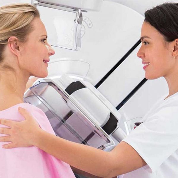
What is Mammography?
Mammography is the low-dose X-ray imaging of the breast tissue to look for early signs of breast cancer before symptoms develop. It can also be used for due diligence when a new symptom (lump or focal pain) develops in the breast tissue.
When viewed on a mammogram, breast tissue appears white and opaque (nebula), while fatty tissue appears darker and translucent.
When Should a Mammogram Be Done?
Annual screening mammograms are recommended for all women from the age of 40. Women who do not have any breast-related signs or symptoms are also screened. If an abnormality is present or patients have a new symptom (a lump or focal pain), additional evaluation may be required. Further examination will reveal what these suspected abnormalities are.
What Percent of Women Who Have Had Mammography Have Risky Situations?
Potential abnormalities are found in 6 to 8 percent of women who get mammograms. This group undergoes different additional evaluations, which may include breast physical examination, diagnostic mammography, breast ultrasound or needle biopsy.After these additional evaluations are completed, it becomes clear what the abnormalities found on the mammogram are.
What Does an Abnormality Look Like on a Mammogram?
The possible abnormality on a mammogram may be called a nodule, mass, lump, density or deterioration:
A mass (lump) with a smooth, well-defined border is usually benign.
Ultrasound is necessary to see and identify the inside of a mass. If the mass contains fluid, it is called a cyst.
A mass (lump) with irregular borders or a starburst appearance may be cancerous and a biopsy is usually recommended.
Microcalcifications (small calcium deposits) are another type of abnormality. They can be classified as benign, suspicious, or uncertain. Most microcalcifications are benign. Depending on how the microcalcifications appear in additional studies (magnification views), a biopsy may be recommended.
What is the Accuracy Rate of Mammography?
Diagnoses made by mammography are between 85 percent and 90 percent accurate. Mammograms can detect breast abnormalities before they are large enough to be felt. However, a palpable mass may not be seen on a mammogram. Any abnormality you feel while examining your breasts should be evaluated by your doctor.
What Should Be Considered Before Mammography?
You can follow your normal routine before the mammogram. You can take your medications and maintain your eating and drinking patterns. If you are breastfeeding, pregnant or think you may be pregnant, you should tell your doctor as your mammogram may need to be postponed.
What Should I Pay Attention to When Coming to My Mammography Appointment?
The technician will ask you to remove one breast from your bib at a time and place it on the chest support plate. The image of the breast is taken by clamping it between two plates. In the meantime, pressure is applied to the breast, preventing the breast from moving. This pressure spreads the breast tissue, allowing the radiologist to see the tissue better. Also, the least amount of radiation is used when the breast is compressed as finely as possible.
You may feel some discomfort during 3-5 seconds of pressure. If you cannot tolerate the pressure, please let the technician know. Pressure can be more bothersome at some times in a woman’s menstrual cycle. To minimize discomfort, we recommend scheduling your appointment seven to 10 days after the start of your period.
How is a Mammogram Taken?
The technician will ask you to remove one breast from your bib at a time and place it on the chest support plate. The image of the breast is taken by clamping it between two plates. In the meantime, pressure is applied to the breast, preventing the breast from moving. This pressure spreads the breast tissue, allowing the radiologist to see the tissue better. Also, the least amount of radiation is used when the breast is compressed as finely as possible.
You may feel some discomfort during 3-5 seconds of pressure. If you cannot tolerate the pressure, please let the technician know. Pressure can be more bothersome at some times in a woman’s menstrual cycle. To minimize discomfort, we recommend scheduling your appointment seven to 10 days after the start of your period.
How Does the Process Proceed After Mammography?
There may be temporary skin discoloration and/or mild pain in the chest due to compression. Most women will be able to resume their normal activities soon after their mammogram. Your results will be available within a few days after the test. After getting the results, your doctor will explain everything to you.
How Often Should You Have a Mammogram?
Regular mammograms every year, starting at the age of 40, will enable you to recognize potential risks early.
What Does the Breast Health Package Consist of?
Breast health package consists of General Surgery Examination and Mammography |
| GENERAL CARDIOLOGY SCREENING PACKAGE |
| LABORATORY ANALYSIS |
| Glucose |
To determine whether or not your blood glucose level is within normal ranges; to screen for, diagnose, and monitor diabetes, and to monitor for the presence of hypoglycaemia (low blood glucose) and hyperglycaemia (high blood glucose) |
| HbA1c |
To monitor average blood glucose levels over a 3 month period. Used to help diagnose and monitor people with diabetes |
| Urea (Bun) |
To measure how much of waste product you have in your blood. It is used to determine how well your kidneys are working |
| Creatinine |
To assess kidney functions |
| Uric Acid |
To diagnose kidney disorder,diagnose and monitor people with gout, monitor kidney function |
| Complete Urinalysis Test |
To look for metabolic and/or kidney disorders and for urinary tract infections |
| Total Cholesterol |
To screen for risk of developing cardiovascular disease (heart disease, stroke and related diseases); to monitor treatment |
| LDL Cholesterol |
| HDL Cholesterol |
| Triglycerides |
| AST (SGOT) |
To diagnose liver, bile duct and heart diseases |
| ALT (SGPT) |
| Sodium |
To investigate causes of dehydration, oedema, problems with blood pressure, or non-specific symptoms |
| Potassium |
To help diagnose and determine the cause of an electrolyte imbalance; to monitor treatment for illnesses that can cause abnormal potassium levels in the body |
| Calcium |
To scan, diagnose, and monitor a range of conditions relating to the bones, heart, nerves, kidneys, and teeth |
| C-Reactive Protein (CRP) |
To identify the presence of inflammation, to determine its severity, and to monitor response to treatment |
| 25 Hydroxy Vitamin D |
To investigate a problem related to bone metabolism or parathyroid function, possible vitamin D deficiency, malabsorption, before commencing specific bone treatment and to monitor some patients taking vitamin D |
| Homocysteine |
To find out if you are at high risk of a heart attack or stroke; also used to determine if you are folate-deficient or vitamin B12-deficient |
| Lipoprotein A |
To evaluate targeted screening for cardiovascular disease (coronary artery disease (CAD) and cerebrovascular disease) risk assessment |
| Blood Count Haemogram |
Haemogram serves as broad screening panel that checks for the presence of any diseases and infections in the body. |
| Troponin |
To see if you have had a heart attack or damage to your heart muscle |
| Vitamin B12 |
To help diagnose the cause of anaemia or neuropathy (nerve damage), to evaluate nutritional status in some
patients, to monitor effectiveness of treatment of B12 or folate deficiency |
| Free T3 |
To help diagnose hyperthyroidism and monitor it's treatment |
| Free T4 |
To diagnose hypothyroidism or hyperthyroidism in adults and to monitor response to treatment |
| TSH |
To screen for and diagnose thyroid disorders; to monitor treatment of hypothyroidism and hyperthyroidism |
| D- dimer |
To help diagnose or exclude thrombotic (blood clot producing) or bleeding diseases and conditions |
| OTHER ANALYSIS |
| Carotid Ultrasound |
To detect narrowing, or stenosis, of the carotid artery, a condition that substantially increases the risk of stroke |
| Echocardiogram |
To evaluate how your heart moves, heart valves are working and heart’s pumping strength |
| Electrocardiogram |
To measure the electrical activity of the heartbeat and hearth rhythm |
| Exercise Stress Test |
To determine how well your hearth handles work. The test can show if the blood supply is reduced in the arteries that supply the heart |
| EXAMINATIONS |
| Cardiology Examination |
General physical examination, evaluation of the results and recommendations |
| General Surgery Examination |
| Dermatology Examination |
|
| Additional information |
| Select Hospital |
Antalya Yaşam Hospital, Kemer Yaşam Hospital, ASV Yaşam Hospital, Opera Yaşam Hospital, Alanya Yaşam Hospital, Manavgat Yaşam Hospital
|
|
| Select Hospital |
Antalya Yaşam Hospital, Kemer Yaşam Hospital, ASV Yaşam Hospital, Opera Yaşam Hospital, Alanya Yaşam Hospital, Manavgat Yaşam Hospital
|
|
| Select Hospital |
Antalya Yaşam Hospital, Kemer Yaşam Hospital, ASV Yaşam Hospital, Opera Yaşam Hospital, Alanya Yaşam Hospital, Manavgat Yaşam Hospital
|
|
| Select Hospital |
Antalya Yaşam Hospital, Kemer Yaşam Hospital, ASV Yaşam Hospital, Opera Yaşam Hospital, Alanya Yaşam Hospital, Manavgat Yaşam Hospital
|
|
| Select Hospital |
Antalya Yaşam Hospital, Kemer Yaşam Hospital, ASV Yaşam Hospital, Opera Yaşam Hospital, Alanya Yaşam Hospital, Manavgat Yaşam Hospital
|
|
| Select Hospital |
Antalya Yaşam Hospital, Kemer Yaşam Hospital, ASV Yaşam Hospital, Opera Yaşam Hospital, Alanya Yaşam Hospital, Manavgat Yaşam Hospital
|
|


Reviews
There are no reviews yet.