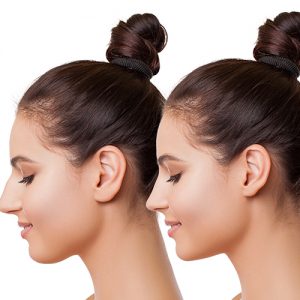
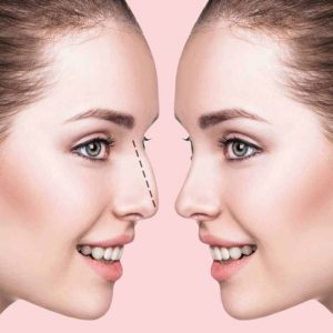
| Content | If you are very close to your ideal nose and there is an uncomfortable appearance only at the tip or holes of the nose, a minor surgical intervention is sufficient with today's technology. With tip plasty, it is possible to experience a change in a short time and easily.
What is Tip plasty?
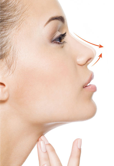
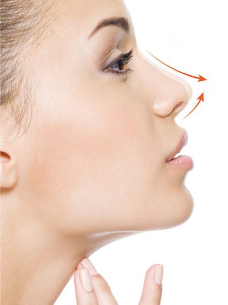
Tip plasty (nasal tip aesthetics) is the surgery performed to give the desired appearance of the cartilage in the front of the nose without touching the nasal bone, in patients who do not have any curvature or deformation in the nasal bone, but have curvature, width, coarseness, irregularity in the nasal anterior cartilage, or wideness in the nostrils.
How long does nose tip plastic surgery take?
The operation takes an average of 20-30 minutes.
Is rhinoplasty a painful operation?
With the drugs given to the nose during the operation, postoperative pain is not felt.
To whom can it be done?
Tip plasty can be performed to beautify the tip of the nose and to eliminate the existing problem in patients who do not have any problems on the back of the nose, the bone structures on the sides of the nose, the septum area in the middle of the nose, the nasal ridge is smooth, the nose is not flattened and wide, and only on the tip of the nose.
With this surgery:
- Thinning the tip of the nose
- Lifting the tip of the nose
- Changing the angle of the nose tip
- Bringing the tip of the nose forward
- Elimination of deformities in the nostrils
- Removal of excess nose wings
- Correction of nasal tip problems can be performed in patients who have had previous nose surgery.
Points to be considered after nasal tip aesthetics and the healing process
Nasal tip aesthetics is a slightly easier surgery with a shorter recovery period compared to other nose surgeries. The patient can return to his daily life in a very short time.
It is recommended to use nasal spray to clean and moisturize the nose after tip plasty. With the cold compress to be applied, the swelling starts to decrease rapidly within 2 days. After the 7th day, the person can easily continue his normal life. The numbness at the tip of the nose may continue for 1 or 2 months. Rapid recovery is achieved in the first 3 months, and full recovery is achieved within 6 to 12 months. |
Vaginoplasty
Vaginoplasty is a procedure to repair the vagina. It treats a variety of medical issues, including vaginal enlargements from childbirth and complications of pelvic floor disease.
What is done during vaginoplasty?
The details of the procedure vary depending on your goals and medical needs. The vagina is reconstructed using various surgical techniques.
Who needs vaginoplasty?
- - Those seeking removal of postpartum enlargement or trauma damage to improve sexual function.
- - Women who need vaginal reconstruction after vaginal exposure to radiation or excision to treat cancer or other conditions.
- - Women who have congenital abnormalities (problems from birth) that affect the development of the vagina.
What is the difference between vaginoplasty and other vaginal operations?
- Vaginoplasty is a surgical procedure to improve the appearance or function of the vagina.Other vaginal procedures:
- - Labiaplasty operation that equalizes or shrinks the labia.
- - A vulvoplasty operation that reshapes the outer part of the vagina.
What is done before vaginoplasty?
- Before the surgery decision, your doctor will perform your physical examination to evaluate your suitability for the operation. During this physical examination, you are expected to provide information about your medical history and general health status.Your doctor will inform you about the risks, benefits and post-operative care requirements of the operation. Listening to your doctor's suggestions and recommendations will reduce the risk of complications.
What is the content of the procedure in women with postpartum deformation?
- In operations performed to correct deformations occurring during childbirth
- - Excess skin is removed,
- - Distorted anatomical lines are repaired,
- - It is ensured that the vaginal opening is reduced.
What is the content of vaginoplasty to repair congenital (birth) defects?
-
- - A functional vagina can be created.
- - Excess tissue or abnormal structures are removed.
- - The structures that cause blood to accumulate in the vagina during menstruation are repaired.
Risks / Benefits
-
- Possible risks of vaginoplasty surgery:
- - Dyspareunia (painful intercourse).
- - It can be summarized as numbness or loss of sensation (usually temporary).
In contrast, the benefits after surgery are:
- - There will be increased sexual satisfaction and self-confidence.
What is the recovery process like after vaginoplasty?
-
- Recovery can take from a few weeks to several months, depending on the extent of the surgery. Your doctor and team will provide you with all the necessary information about post-operative care after vaginoplasty.
How are the controls planned after vaginoplasty?
-
- Keeping in touch with your doctor after the surgery and not interrupting your controls will speed up your recovery process and minimize the risk of complications.
|
| MEN UNDER 40 LARGE SCREENING PACKAGE |
| Glucose |
To determine whether or not your blood glucose level is within normal ranges; to screen for, diagnose, and monitor diabetes, and to monitor for the presence of hypoglycaemia (low blood glucose) and hyperglycaemia (high blood glucose) |
| HbA1c |
To monitor average blood glucose levels over a 3 month period. Used to help diagnose and monitor people with diabetes |
| Urea (Bun) |
To measure how much of waste product you have in your blood. It is used to determine how well your kidneys are working |
| Creatinine |
To assess kidney functions |
| Uric Acid |
To diagnose kidney disorder,diagnose and monitor people with gout, monitor kidney function |
| Complete Urinalysis Test |
To look for metabolic and/or kidney disorders and for urinary tract infections |
| Total Cholesterol |
To screen for risk of developing cardiovascular disease (heart disease, stroke and related diseases); to monitor treatment |
| LDL Cholesterol |
| HDL Cholesterol |
| Triglycerides |
| AST (SGOT) |
To diagnose liver, bile duct and heart diseases |
| ALT (SGPT) |
| GGT |
To screen for liver disease or alcohol abuse; and to help your doctor tell whether a raised concentration of alkaline phosphatase (ALP) in the bloodstream is due to liver or bone disease |
| ALP |
To screen for or monitor treatment for liver or bone disorder |
| Sodium |
To investigate causes of dehydration, oedema, problems with blood pressure, or non-specific symptoms |
| Potassium |
To help diagnose and determine the cause of an electrolyte imbalance; to monitor treatment for illnesses that can cause abnormal potassium levels in the body |
| Chloride |
To determine if there is a problem with your body’s acid-alkali (pH) balance and to monitor treatment |
| Calcium |
To scan, diagnose, and monitor a range of conditions relating to the bones, heart, nerves, kidneys, and teeth. |
| Phosphate |
To help in the diagnosis of conditions known to cause abnormally high or low levels |
| Amylase |
To diagnose pancreatitis or other pancreatic diseases |
| Lipase |
To diagnose and monitor pancreatitis or other pancreatic disease |
| Magnesium |
To measure the concentration of magnesium in your blood and to help determine the cause of abnormal calcium and/or potassium levels |
| C-Reactive Protein (CRP) |
To identify the presence of inflammation, to determine its severity, and to monitor response to treatment |
| 25 Hydroxy Vitamin D |
To investigate a problem related to bone metabolism or parathyroid function, possible vitamin D deficiency, malabsorption, before commencing specific bone treatment and to monitor some patients taking vitamin D |
| Blood Count Haemogram |
Haemogram serves as broad screening panel that checks for the presence of any diseases and infections in the body |
| Erythrocyte Sedimentation Rate
(ESR) |
To detect and monitor the activity of inflammation as an aid in the diagnosis of the underlying cause |
| Ferritine |
To help assess the levels of iron stored in your body |
| Vitamin B12 |
To help diagnose the cause of anaemia or neuropathy (nerve damage), to evaluate nutritional status in some patients, to monitor effectiveness of treatment of B12 or folate deficiency |
| Free T3 |
To help diagnose hyperthyroidism and monitor it's treatment |
| Free T4 |
To diagnose hypothyroidism or hyperthyroidism in adults and to monitor response to treatment |
| TSH |
To screen for and diagnose thyroid disorders; to monitor treatment of hypothyroidism and hyperthyroidism |
| HBsAg |
To detect, diagnose and follow the course of an infection with hepatitis B virus (HBV) or to determine if the vaccine against hepatitis B has produced the desired level of immunity |
| Anti HBs |
| Anti HCV |
To screen for and diagnose hepatitis C virus infection and to monitor treatment of the infection |
| Anti HIV |
To determine if you are infected with human immunodeficiency virus (HIV) |
| Fecal Occult Blood Test |
To screen for bleeding from the gut/intestine, which may be an indicator of bowel cancer |
| OTHER ANALYSIS |
| Abdominal Ultrasound |
To identify diseases at organs in the abdomen, including the liver, gallbladder, spleen, pancreas, and kidneys |
| Thyroid Ultrasound |
To characterize a thyroid nodule(s), i.e. to measure the dimensions accurately and to identify internal structure and vascularization |
| Echocardiogram |
To evaluate how your heart moves, heart valves are working and heart’s pumping strength |
| Electrocardiogram |
To measure the electrical activity of the heartbeat and hearth rhythm |
| Exercise Stress Test |
To determine how well your hearth handles work. The test can show if the blood supply is reduced in the arteries that supply the heart |
| Pulmonary Function Test |
To tests that measure how well your lungs work. |
| Chest X-Ray |
The most commonly preferred diagnostic examination to produce images of heart, lungs, airways, blood vessels and the bones of the spine and chest |
| EXAMINATIONS |
| Internal Medicine Examination |
General physical examination, evaluation of the results and recommendations |
| Cardiology Examination |
| Ophtalmology Examination |
| Pulmonology Examination |
| Urology Examination |
| General Surgery Examination |
| Dermatology Examination |
| Breast reduction surgery is an operation performed to bring the breasts that are larger than the person's body to normal sizes. |
What is laser tattoo removal?
Tattooing is a permanent procedure on the skin. Tattoo removal can be done with modern methods when, for any reason, the person does not want to see his tattoo on his body anymore.
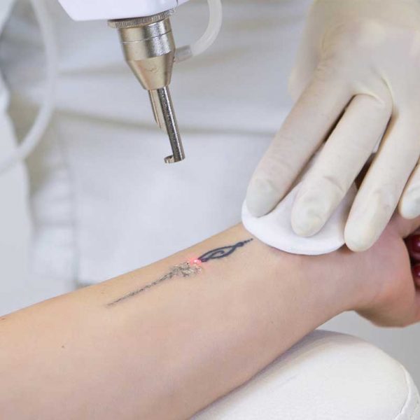
Tattoo removal, permanent make-up and stain treatment can be performed with the Q-switch Nd:YAG laser. This process is a technology that destroys the color pigments used in tattoos by focusing. During this very delicate process, the laser sees and destroys the pigment and damages the tissue as little as possible. In laser tattoo removal, which is a long and laborious process, one hundred percent result may not always be achieved. For this, the decision to have a tattoo should be considered very well, and possible future regrets should be considered.
How is laser tattoo removal done?
Tattoo and skin analysis should be done before the procedure. Lasers designed for tattoo removal and spot treatment send the beam to the epidermis layer of the skin, absorb the compounds that give color to the dermis and subcutaneous tattoo, and destroy the chemical substance by breaking it down. The wavelength of the laser; is adjusted according to the size, density and color of the tattoo.
In laser shots, dye pigments are made without damaging the tissues.
While black dyes under the skin are better detected by the laser, more effective results are obtained, while the same is not true for light-colored dyes.
Different wave sizes are used in tattoos with multi-colored paints.
How many sessions does laser tattoo removal take?
There are some environmental and structural factors in tattoo removal. It is important in which part of the body the tattoo is located. More sessions are required to remove tattoos, especially on the tips of the feet and fingers. For tattoos on larger areas of the body, a more effective process is performed and removed. The duration of the erasing process varies due to the color of the tattoo, the amount and type of paint used in the tattoo. Tattoo removal sessions take place at intervals of approximately 1.5 – 2 months. The number of sessions varies according to the person and the nature of the tattoo.
Will there be any scars after laser tattoo removal?
After each tattoo removal session, the color of the tattoo will lighten by approximately 20%. In tattoos where multi-colored and phosphorescent colors are used, the erasing process may be longer.
Is laser tattoo removal painful?
After numbing the skin part with cream or local anesthesia, the procedure is performed by protecting the upper skin layer in that area with the help of a Q-Switch ND-YAG laser and cooler. After the procedure, there may be bleeding in the area and crusting on the skin that will last for about a few days after the procedure. After the application, antibiotic cream should be used to both repair the area and prevent the development of infection.
When should laser tattoo removal be done?
While tattoo removal is more comfortable in winter, it is not preferred in summer due to the risk of staining with the effect of the sun. The time of the procedure varies according to the size of the tattoo. Since the sun is out in the summer, problems such as permanent staining may occur after the procedure. The best time for this is the winter months. |
| WOMEN OVER 40 LARGE SCREENING PACKAGE |
| LABORATORY ANALYSIS |
| Glucose |
To determine whether or not your blood glucose level is within normal ranges; to screen for, diagnose, and monitor diabetes, and to monitor for the presence of hypoglycaemia (low blood glucose) and hyperglycaemia (high blood glucose) |
| HbA1c |
To monitor average blood glucose levels over a 3 month period. Used to help diagnose and monitor people with diabetes |
| Urea (Bun) |
To measure how much of waste product you have in your blood. It is used to determine how well your kidneys are working |
| Creatinine |
To assess kidney functions |
| Uric Acid |
To diagnose kidney disorder,diagnose and monitor people with gout, monitor kidney function |
| Complete Urinalysis Test |
To look for metabolic and/or kidney disorders and for urinary tract infections |
| Total Cholesterol |
To screen for risk of developing cardiovascular disease (heart disease, stroke and related diseases); to monitor treatment |
| LDL Cholesterol |
| HDL Cholesterol |
| Triglycerides |
| AST (SGOT) |
To diagnose liver, bile duct and heart diseases |
| ALT (SGPT) |
| GGT |
To screen for liver disease or alcohol abuse; and to help your doctor tell whether a raised concentration of alkaline phosphatase (ALP) in the bloodstream is due to liver or bone disease |
| ALP |
To screen for or monitor treatment for liver or bone disorder |
| Sodium |
To investigate causes of dehydration, oedema, problems with blood pressure, or non-specific symptoms |
| Potassium |
To help diagnose and determine the cause of an electrolyte imbalance; to monitor treatment for illnesses that can cause abnormal potassium levels in the body |
| Chloride |
To determine if there is a problem with your body’s acid-alkali (pH) balance and to monitor treatment |
| Calcium |
To scan, diagnose, and monitor a range of conditions relating to the bones, heart, nerves, kidneys, and teeth |
| Phosphate |
To help in the diagnosis of conditions known to cause abnormally high or low levels |
| Amylase |
To diagnose pancreatitis or other pancreatic diseases |
| Lipase |
To diagnose and monitor pancreatitis or other pancreatic disease |
| Magnesium |
To measure the concentration of magnesium in your blood and to help determine the cause of abnormal calcium and/or potassium levels |
| C-Reactive Protein (CRP) |
To identify the presence of inflammation, to determine its severity, and to monitor response to treatment |
| 25 Hydroxy Vitamin D |
To investigate a problem related to bone metabolism or parathyroid function, possible vitamin D deficiency, malabsorption, before commencing specific bone treatment and to monitor some patients taking vitamin D |
| Blood Count Haemogram |
Haemogram serves as broad screening panel that checks for the presence of any diseases and infections in the body |
| Erythrocyte Sedimentation Rate
(ESR) |
To detect and monitor the activity of inflammation as an aid in the diagnosis of the underlying cause |
| Ferritine |
To help assess the levels of iron stored in your body |
| Vitamin B12 |
To help diagnose the cause of anaemia or neuropathy (nerve damage), to evaluate nutritional status in some patients, to monitor effectiveness of treatment of B12 or folate deficiency |
| Free T3 |
To help diagnose hyperthyroidism and monitor it's treatment |
| Free T4 |
To diagnose hypothyroidism or hyperthyroidism in adults and to monitor response to treatment |
| TSH |
To screen for and diagnose thyroid disorders; to monitor treatment of hypothyroidism and hyperthyroidism |
| FSH |
To evaluate the function of your pituitary gland, which regulates the hormones that control your reproductive system |
| LH |
| Estrogens (E2) |
To measure or monitor your estrogen levels; to detect an abnormal level or hormone imbalance ; to monitor treatment for infertility or symptoms of menopause; sometimes to test for fetal-placental status during early stages of pregnancy |
| Prolactin |
To diagnose a prolactinoma (a type of tumor of the pituitary gland, help tofind the cause of a woman's menstrual irregularities and/or infertility. Or to help to find the cause of a man's low sex drive and/or erectile dysfunction |
| Beta hCG |
To confirm and monitor pregnancy or to diagnose trophoblastic disease or germ cell tumours |
| HBsAg |
To detect, diagnose and follow the course of an infection with hepatitis B virus (HBV) or to determine if the vaccine against hepatitis B has produced the desired level of immunity |
| Anti HBs |
| Anti HCV |
To screen for and diagnose hepatitis C virus infection and to monitor treatment of the infection |
| Anti HIV |
To determine if you are infected with human immunodeficiency virus (HIV) |
| CEA |
In the presence of certain cancers, CEA may be used to monitor the effect of treatment and recurrence of disease |
| CA 125 |
To monitor treatment for ovarian cancer or to investigate for a possible ovarian cancer |
| CA 19-9 |
To help tell the difference between cancer of the pancreas and bile ducts and other conditions; to monitor response to pancreatic cancer treatment and to watch for recurrence |
| CA 15-3 |
To monitor the response to treatment of breast cancer and to watch for recurrence of the disease |
| AFP |
To screen for and monitor therapy for certain cancers of the liver and testes |
| Fecal Direct Parasite Search (Ova
& Parasite Exam) |
To determine whether you have a parasite infecting your digestive tract |
| Fecal Occult Blood Test |
To screen for bleeding from the gut/intestine, which may be an indicator of bowel cancer |
| OTHER ANALYSIS |
| Abdominal Ultrasound |
To identify diseases at organs in the abdomen, including the liver, gallbladder, spleen, pancreas, and kidneys |
| Thyroid Ultrasound |
To characterize a thyroid nodule(s), i.e. to measure the dimensions accurately and to identify internal structure and vascularization |
| Carotid Ultrasound |
To detect narrowing, or stenosis, of the carotid artery, a condition that substantially increases the risk of stroke |
| Breast Ultrasound (Bilateral) |
To screen suspected breast cancer or for early diagnosis and control. It is the imaging of breast with ultrasound device |
| Mammography (Bilateral) |
| Pap Smear |
Method for early diagnosis of Cervical cancer and infectious diseases |
| Echocardiogram |
To evaluate how your heart moves, heart valves are working and heart’s pumping strength |
| Electrocardiogram |
To measure the electrical activity of the heartbeat and hearth rhythm |
| Exercise Stress Test |
To determine how well your hearth handles work. The test can show if the blood supply is reduced in the arteries that supply the heart |
| Pulmonary Function Test |
To tests that measure how well your lungs work. |
| Chest X-Ray |
The most commonly preferred diagnostic examination to produce images of heart, lungs, airways, blood vessels and the bones of the spine and chest |
| EXAMINATIONS |
| Internal Medicine Examination |
General physical examination, evaluation of the results and recommendations |
| Cardiology Examination |
| Gynaecology Examination |
| Ophtalmology Examination |
| Pulmonology Examination |
| General Surgery Examination |
|
|
|













Reviews
There are no reviews yet.