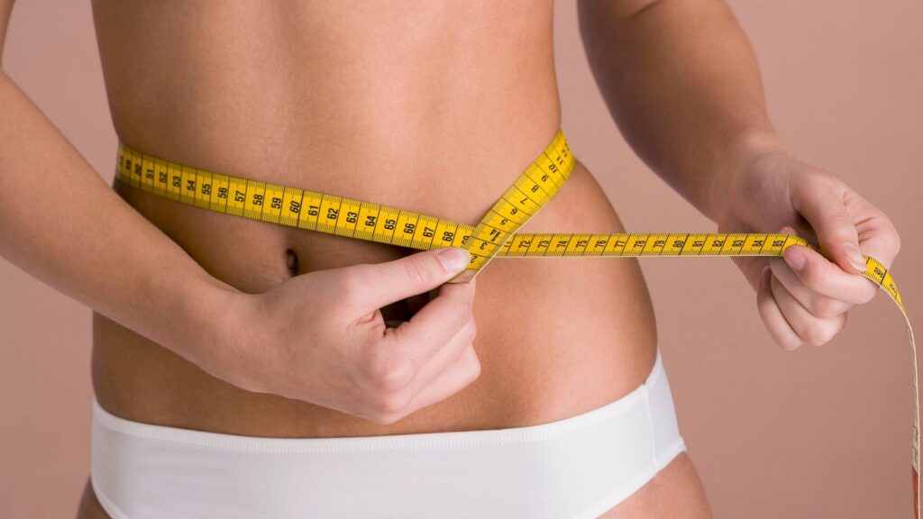
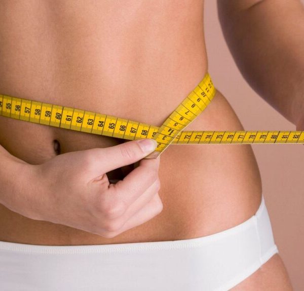
| Content | A mini tummy tuck is a good option for patients who have excess weight in the abdomen, whose skin is too loose for liposuction to be performed alone, but who is too tight to require a full tummy tuck. A mini tummy tuck is a frequently preferred method in patients who are not overweight, as it causes shorter scars and faster post-operative recovery.
What is a mini tummy tuck (mini-abdominoplasty)?
In the mini tummy tuck process, excess fat is removed from the entire abdomen and waist area, excess skin under the navel is removed, but no stretching is performed on the part above the navel.
Who can have a mini tummy tuck?
Mini tummy tuck is applied to patients who have given birth but whose skin and excess fat are limited below the navel. Patients with a high navel are also suitable candidates for a mini tummy tuck.
How are mini tummy tuck surgeries performed?
In the mini tummy tuck procedure, liposuction is first applied to the entire abdomen and waist area. In the meantime, if necessary, fat injection is made into the buttocks. The abdominal skin is lifted from the abdominal wall up to the navel. If necessary, the loosened abdominal muscles are stitched together and the lower part of the abdominal wall is tightened. Then, the excess part of the skin under the belly button is removed.
Is it necessary to lose weight before mini tummy tuck surgery?
Excess weight poses a risk to all surgeries. After the operation, patients stand up more difficult, their wounds heal later, and lung problems are experienced more frequently. For this reason, obese patients are recommended to reduce their weight before surgery.
Is anesthesia required in mini tummy tuck surgeries?
Abdominoplasty operations are performed in the operating room environment and under general anesthesia or heavy sedatives.
How long does tummy tuck surgery take?
The procedures to be performed determine the duration of the surgery. If liposuction will not be applied to a very large area, mini tummy tuck surgery takes between one and two hours.
What happens after mini tummy tuck surgery?
During the operation, a drain is often placed on the patients and an abdominal corset is worn. The patient is fed and stood up three to four hours after surgery. Usually the patient is discharged the same or the next day. If necessary, the drains are left in place for a few more days. Since all the sutures are left under the skin, there is no need for suture removal. Two to three days after surgery, patients can go back to their normal life. After six weeks, all sports activities are free. |
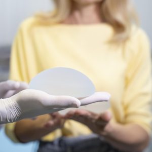
Breast Health Package for Women Under 40
Our Breast Health Center, which is a part of Yaşam Hospital Oncology Center, offers all the possibilities of technology to provide the best care to every woman.
Early Detection of Breast Cancer
Breast cancer is the most common type of cancer that women can encounter in their lifetime (Early detection can save your life).
Self Examination
From the age of 20, all women should perform a breast self-exam once a month (about one week after the start of each menstrual period). If you feel an unusual lump or anything else, it’s important to contact your doctor right away.
What is Breast Ultrasound?
Ultrasound is an imaging method that uses high-frequency sound waves to take pictures of internal organs and tissues.
When Should Breast Ultrasound Be Done?
From the age of 25, annual routine ultrasound follow-ups should not be interrupted. Ultrasound can be applied safely as it does not contain radiation and has no side effects on the body.
What Are the Risks of Breast Ultrasound?
Breast ultrasound uses sound waves, not radiation, to produce images. Ultrasound technology has no known risks.
What Happens After Breast Ultrasound?
After your breast ultrasound, the radiologist interprets the images and reports the results to your doctor. This information will also be shared with you if any additional testing is needed or follow-up is recommended.
What are the Benefits of Breast Ultrasound?
Ultrasound scanning is non-invasive (no needles or injections).
Ultrasound imaging is extremely safe and uses no radiation.
An ultrasound scan gives a clear picture of soft tissues that don’t show up well on x-ray images.
Ultrasound provides real-time imaging. This makes it easy to guide minimally invasive procedures such as needle biopsies and fluid aspiration.
Ultrasound imaging helps detect lesions in women with dense breasts.
Ultrasound can help detect and classify a breast lesion that cannot be adequately interpreted by mammography alone.
Using ultrasound, doctors can determine that many areas are caused by normal tissue (such as fat lobules) or benign cysts. For most women age 40 and older, a mammogram will be used along with the ultrasound. For women under the age of 40, ultrasound alone is often sufficient to determine whether an area of concern needs a biopsy.
What Does the Breast Health Package Consist of?
Breast health package consists of General Surgery Examination and Breast USG.
Note from your doctor:
Breast ultrasound is a safe, painless imaging method for examining targeted areas of breast tissue. With breast ultrasound, we provide detailed images of breast tissue and can diagnose cysts or lumps if present. Diagnosing possible risks at an early stage provides a very important gain in the treatment process. | Breast reduction surgery is an operation performed to bring the breasts that are larger than the person's body to normal sizes. |
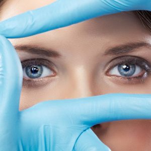
Labiaplasty Package
Labiaplasty is a surgical procedure to reduce or enlarge the skin folds that usually surround your labia and vagina. Excess skin can be bent and pinched, causing discomfort during exercise, physical activities, and sexual intercourse.
Depending on why the procedure is performed during labiaplasty, your doctor may:
It removes some tissue from the labia to reduce its size.
Injects filler or oil to enlarge the labia.
It forms a labia from other tissues.
What are the reasons for requesting labiaplasty?
Reducing labia minora size
Excess lip tissue can be bent, twisted, compressed. This, can cause physical discomfort and irritation during exercise, physical activities (such as cycling or jogging), and sexual intercourse.
Reducing the size of the labia minora may be desirable to improve hygiene and health, as excess tissue can make cleaning difficult and may harbor bacteria that can lead to the development of urinary tract infections.
Cosmetic and emotional reasons
To have a younger appearance after birth or aging,
Reducing asymmetry of the labia minora or labia majora when one side is longer or has a different shape than the other,
To increase self-confidence and to eliminate visual lines and protrusions when wearing body-fitting trousers or tights,
During close contact, labiaplasty can be performed to improve comfort, confidence and sexual health regarding the appearance of your genitals.
What causes an overgrowth of a labia?
Age, menopause, or other hormonal changes can thin the labia majora tissue, causing the labia minora to protrude from the labia majora.
Pregnancy and childbirth.
Changes in your weight.
Genetic.
What are the pre-operative procedures?
First, you and your doctor will discuss why you want to have labiaplasty and make sure that the surgery will meet your expectations. During the physical examination, your doctor will explain where the incisions will be made and what to expect regarding changes in the size and shape of your labia.
After the surgery decision, your doctor will inform you in detail about the preparation processes for the surgery. In this process, your doctor will inform you about how to resize or reshape your labia (labia majora and/or labia minora) and will enlighten you about the surgical procedure to be applied.
What is the procedure after labiaplasty surgery?
After the surgery, your doctor will give you the necessary information on how to care. Carefully applying the information conveyed to you about what you should and shouldn’t do during the recovery process will speed up your recovery process. We also recommend that you make your control appointments before leaving the hospital.
What are the risks and complications of labiaplasty surgery?
- - Complications of laboplasty surgery are rare. Even a little;
- - Bleeding,
- - Bruising, swelling (hematoma),
- - Infection,
- - Scar,
- - Ongoing pain, pain with sexual intercourse, or loss of sensitivity may occur.
- - In these cases, you can consult your doctor.
Is the recovery process painful after labiaplasty?
- It is normal to feel some swelling, discomfort and pain after surgery. Most people report only mild pain or tenderness for a few days. In return, your doctor will advise you on medication for your pain.Wearing loose trousers and underwear during the recovery period will accelerate your healing process.
Will it bleed a lot?
You may have a small amount of bleeding for a week. This is normal, you can use pads to absorb the blood.
When will the satisfactory results of labiaplasty be obtained?
Initial swelling, pain, and temporary discoloration subside within a few weeks after surgery. However, you may have to wait up to four to six months to see the final results of your labiaplasty. Post-operative scarring is usually negligible.
How do I wash my labia area?
Use only warm water to wash your wound (do not use soap) and wipe gently, do not rub the surgical site.
It is important to follow your doctor’s instructions to keep the surgical site clean and free of bacteria and to speed the healing of the surgical site.
When can I return to work, physical activity and sex life?
You should be able to return to work and other light activities after a few days. If your job involves intense physical activity or lifting weights, you can get more detailed information by informing your doctor about this. until your doctor approves. |
| OVERALL CARDIOLOGY SCREENING PACKAGE |
| LABORATORY ANALYSIS
|
| Glucose |
To determine whether or not your blood glucose level is within normal ranges; to screen for, diagnose, and monitor diabetes, and to monitor for the presence of hypoglycaemia (low blood glucose) and hyperglycaemia (high blood glucose) |
| HbA1c |
To monitor average blood glucose levels over a 3 month period. Used to help diagnose and monitor people with diabetes. |
| Urea (Bun) |
To measure how much of waste product you have in your blood. It is used to determine how well your kidneys are working |
| Creatinine |
To assess kidney functions |
| Uric Acid |
To diagnose kidney disorder,diagnose and monitor people with gout, monitor kidney function. |
| Complete Urinalysis Test |
To look for metabolic and/or kidney disorders and for urinary tract infections |
| Total Cholesterol |
To screen for risk of developing cardiovascular disease (heart disease, stroke and related diseases); to monitor treatment |
| LDL Cholesterol |
| HDL Cholesterol |
| Triglycerides |
| AST (SGOT) |
To diagnose liver, bile duct and heart diseases. |
| ALT (SGPT) |
| Sodium |
To investigate causes of dehydration, oedema, problems with blood pressure, or non-specific symptoms |
| Potassium |
To help diagnose and determine the cause of an electrolyte imbalance; to monitor treatment for illnesses that can cause abnormal potassium levels in the body |
| Calcium |
To scan, diagnose, and monitor a range of conditions relating to the bones, heart, nerves, kidneys, and teeth |
| C-Reactive Protein (CRP) |
To identify the presence of inflammation, to determine its severity, and to monitor response to treatment |
| 25 Hydroxy Vitamin D |
To investigate a problem related to bone metabolism or parathyroid function, possible vitamin D deficiency, malabsorption, before commencing specific bone treatment and to monitor some patients taking vitamin D |
| Homocysteine |
To find out if you are at high risk of a heart attack or stroke; also used to determine if you are folate-deficient or vitamin B12-deficient |
| Lipoprotein A |
To evaluate targeted screening for cardiovascular disease (coronary artery disease (CAD) and cerebrovascular disease) risk assessment |
| Blood Count Haemogram |
Haemogram serves as broad screening panel that checks for the presence of any diseases and infections in the body. |
| Troponin |
To see if you have had a heart attack or damage to your heart muscle |
| Vitamin B12 |
To help diagnose the cause of anaemia or neuropathy (nerve damage), to evaluate nutritional status in some patients, to monitor effectiveness of treatment of B12 or folate deficiency |
| Free T3 |
To help diagnose hyperthyroidism and monitor it's treatment |
| Free T4 |
To diagnose hypothyroidism or hyperthyroidism in adults and to monitor response to treatment |
| TSH |
To screen for and diagnose thyroid disorders; to monitor treatment of hypothyroidism and hyperthyroidism |
| D- dimer |
To help diagnose or exclude thrombotic (blood clot producing) or bleeding diseases and conditions |
| OTHER ANALYSIS |
| Carotid Ultrasound |
To detect narrowing, or stenosis, of the carotid artery, a condition that substantially increases the risk of stroke |
| Echocardiogram |
To evaluate how your heart moves, heart valves are working and heart’s pumping strength |
| Electrocardiogram |
To measure the electrical activity of the heartbeat and hearth rhythm |
| Coronary CT Angiography |
It is a imaging method performed by computed tomography of the coronary arteries, which are the vessels that feed the heart, by giving contrast material through the vein of the forearm |
| EXAMINATIONS |
| Cardiology Examination |
General physical examination, evaluation of the results and recommendations |
| General Surgery Examination |
| Dermatology Examination |
|
| GENERAL CARDIOLOGY SCREENING PACKAGE |
| LABORATORY ANALYSIS |
| Glucose |
To determine whether or not your blood glucose level is within normal ranges; to screen for, diagnose, and monitor diabetes, and to monitor for the presence of hypoglycaemia (low blood glucose) and hyperglycaemia (high blood glucose) |
| HbA1c |
To monitor average blood glucose levels over a 3 month period. Used to help diagnose and monitor people with diabetes |
| Urea (Bun) |
To measure how much of waste product you have in your blood. It is used to determine how well your kidneys are working |
| Creatinine |
To assess kidney functions |
| Uric Acid |
To diagnose kidney disorder,diagnose and monitor people with gout, monitor kidney function |
| Complete Urinalysis Test |
To look for metabolic and/or kidney disorders and for urinary tract infections |
| Total Cholesterol |
To screen for risk of developing cardiovascular disease (heart disease, stroke and related diseases); to monitor treatment |
| LDL Cholesterol |
| HDL Cholesterol |
| Triglycerides |
| AST (SGOT) |
To diagnose liver, bile duct and heart diseases |
| ALT (SGPT) |
| Sodium |
To investigate causes of dehydration, oedema, problems with blood pressure, or non-specific symptoms |
| Potassium |
To help diagnose and determine the cause of an electrolyte imbalance; to monitor treatment for illnesses that can cause abnormal potassium levels in the body |
| Calcium |
To scan, diagnose, and monitor a range of conditions relating to the bones, heart, nerves, kidneys, and teeth |
| C-Reactive Protein (CRP) |
To identify the presence of inflammation, to determine its severity, and to monitor response to treatment |
| 25 Hydroxy Vitamin D |
To investigate a problem related to bone metabolism or parathyroid function, possible vitamin D deficiency, malabsorption, before commencing specific bone treatment and to monitor some patients taking vitamin D |
| Homocysteine |
To find out if you are at high risk of a heart attack or stroke; also used to determine if you are folate-deficient or vitamin B12-deficient |
| Lipoprotein A |
To evaluate targeted screening for cardiovascular disease (coronary artery disease (CAD) and cerebrovascular disease) risk assessment |
| Blood Count Haemogram |
Haemogram serves as broad screening panel that checks for the presence of any diseases and infections in the body. |
| Troponin |
To see if you have had a heart attack or damage to your heart muscle |
| Vitamin B12 |
To help diagnose the cause of anaemia or neuropathy (nerve damage), to evaluate nutritional status in some
patients, to monitor effectiveness of treatment of B12 or folate deficiency |
| Free T3 |
To help diagnose hyperthyroidism and monitor it's treatment |
| Free T4 |
To diagnose hypothyroidism or hyperthyroidism in adults and to monitor response to treatment |
| TSH |
To screen for and diagnose thyroid disorders; to monitor treatment of hypothyroidism and hyperthyroidism |
| D- dimer |
To help diagnose or exclude thrombotic (blood clot producing) or bleeding diseases and conditions |
| OTHER ANALYSIS |
| Carotid Ultrasound |
To detect narrowing, or stenosis, of the carotid artery, a condition that substantially increases the risk of stroke |
| Echocardiogram |
To evaluate how your heart moves, heart valves are working and heart’s pumping strength |
| Electrocardiogram |
To measure the electrical activity of the heartbeat and hearth rhythm |
| Exercise Stress Test |
To determine how well your hearth handles work. The test can show if the blood supply is reduced in the arteries that supply the heart |
| EXAMINATIONS |
| Cardiology Examination |
General physical examination, evaluation of the results and recommendations |
| General Surgery Examination |
| Dermatology Examination |
|


Reviews
There are no reviews yet.